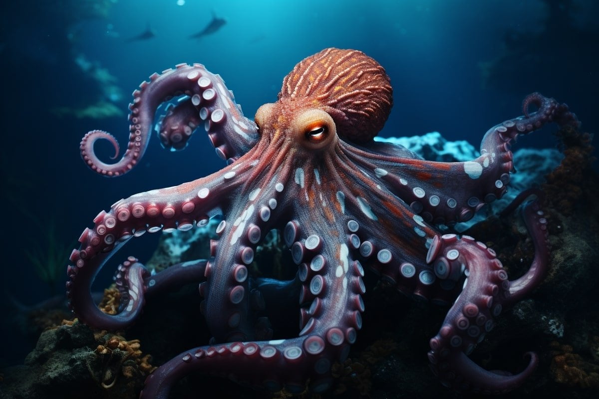
summary: The researchers mapped neural activity in the octopus’s visual system, revealing striking similarities with humans.
The team observed neural responses to the bright and dark dots, thus mapping what resembles the organization of the human brain. Interestingly, octopuses and humans shared the last common ancestor around 500 million years ago, suggesting an independent evolution of such complex visual systems.
These findings contribute significantly to our understanding of cephalopod vision and brain structure.
Key facts:
- About 70% of an octopus’s brain is dedicated to vision. This research is the first of its kind to map neural activity in their visual system, providing insights into how these marine creatures perceive their world.
- Despite having a common ancestor 500 million years ago, octopuses and humans evolved similar neural maps for visual perception.
- The study discovered that octopus neurons respond strongly to small light spots and large dark spots, which differ from the human visual system. This is likely due to the peculiarities of the underwater environment.
source: University of Oregon
An octopus devotes about 70 percent of its brain to vision. But until recently, scientists had only a vague understanding of how these marine animals see their underwater world. A new University of Oregon study sheds light on the octopus’s point of view.
For the first time, neuroscientists have recorded neural activity from an octopus’ visual system. They created a map of the octopus’ visual field by directly observing neural activity in the animal’s brain in response to light and dark spots at different locations.
This map of neural activity in an octopus’s visual system is very similar to what we see in a human brain — even though octopuses and humans shared a common ancestor some 500 million years ago, octopuses evolved their complex nervous systems independently.
Neuroscientist Christopher Neale and his team report their findings in a paper published June 20 in the journal Neuroscientist Christopher Neale. Current Biology.
“Nobody’s actually recorded from the central visual system of a cephalopod before,” Neal said. Octopuses and other cephalopods aren’t usually used as models for understanding vision, but Neal’s team is intrigued by their unusual brains.
In a related paper published last year in Current BiologyThe lab identified different classes of neurons in an octopus’ optic lobe, a part of the brain dedicated to vision. “Together, these papers provide a good foundation by showing the different types of neurons and what they respond to—two key aspects we want to know to start understanding a new visual system,” Neal said.
In the new study, the researchers measured how neurons in the octopus’ visual system respond to dark and light spots moving across a screen. Using fluorescent microscopy, researchers can watch the activity of neurons as they respond, to see how neurons react differently depending on where the spots appear.
“We were able to see that each site in the optic lobe responded to a single location on the screen in front of the animal,” Neal said. “If we move somewhere, the response moves in the brain.”
This type of individual maps are found in the human brain for multiple senses, such as vision and touch. Neuroscientists have linked the location of certain sensations to specific spots in the brain.
A well-known representation of touch is the homunculus, a cartoon human figure in which the parts of the body are drawn in proportion to the amount of brain space devoted to processing sensory input there.
Highly sensitive spots such as fingers and toes appear huge because there is a lot of brain input from these body parts, while less sensitive areas are much smaller.
But finding an orderly connection between the visual scene and the octopus brain was far from the case. It is a rather complex evolutionary innovation, and some animals such as reptiles do not have this type of map. Also, previous studies have indicated that octopuses do not have a homunculus-like map of different parts of their bodies.
“We hoped the visual map was there, but no one had directly noticed it before,” Neal said.
The researchers also noted that neurons in the octopus responded particularly strongly to small light spots and large dark spots – a marked difference from the human visual system. Neal’s team hypothesizes that this may be due to specific characteristics of the underwater environment that the octopuses must navigate. Looming predators may appear as large dark shadows, while nearby objects such as food may appear as small bright spots.
Next, the researchers hope to understand how the octopus’s brain responds to more complex images, such as those already in their natural environment. Their ultimate goal is to trace the pathway of these visual inputs deeper into the octopus’ brain, to understand how the octopus sees and interacts with its world.
About this research in Visual Neuroscience News
author: Molly Blancett
source: University of Oregon
communication: Molly Blancett – University of Oregon
picture: Image credited to Neuroscience News
Original search: open access.
“Functional regulation of visual responses in the optic lobe of an octopusWritten by Christopher Neal, et al. Current Biology
a summary
Functional regulation of visual responses in the optic lobe of an octopus
Highlights
- The functional organization of the cephalopod visual system is largely unknown
- Using calcium imaging, we mapped visual responses in the octopus’ optic lobe
- We identified spatially localized receptive fields with retinal organization
- The on and off pathways were distinct and had unique size-selective properties
summary
Cephalopods are highly visual animals with camera-type eyes, large brains, and a rich repertoire of visually-directed behaviors. However, the brain of cephalopods evolved independently of the brain of other species with high vision, such as vertebrates. Therefore, the neural circuits that process sensory information are very different.
It is largely unknown how their uniquely powerful visual system works, as there have been no direct neurological measurements of visual responses in the cephalopod brain.
In this study, we used two-photon calcium imaging to record visually evoked responses in the primary visual processing center of the octopus central brain, the optic lobe, to determine how basic features of the visual scene are represented and organized.
We found spatially localized receptive domains of light (ON) and dark (OFF) stimuli, which were retinally organized across the optic lobe, demonstrating the hallmark of visual system organization common across many species.
Examination of these responses revealed shifts in visual representation across layers of the visual lobe, including emergence of the OFF pathway and increased size selectivity.
We also identified asymmetries in the spatial processing of on and off stimuli, which suggest unique circuit mechanisms for model processing that may have evolved to accommodate the specific requirements of processing an underwater visual scene.
This study provides insights into the neural processing and functional organization of the octopus visual system, highlighting both common and unique aspects, and lays a foundation for future studies of the neural circuits mediating visual processing and behavior in cephalopods.

“Web maven. Infuriatingly humble beer geek. Bacon fanatic. Typical creator. Music expert.”

:quality(85)/cloudfront-us-east-1.images.arcpublishing.com/infobae/LKGZZPZZ6THIZNWF5WHDNKDV64.jpg)


More Stories
NASA Commercial Crew Comparison Boeing Starliner and SpaceX Dragon
Japanese “Moon Sniper” brings back images after the third long lunar night
Weather web maps on the exoplanet WASP-43b